| ||||||||||||||||||||


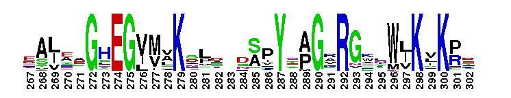
 Adenylation domain of archaeal and bacterial LigB-like DNA ligases. ATP-dependent polynucleotide ligases catalyze phosphodiester bond formation using nicked nucleic acid substrates with the high energy nucleotide of ATP as a cofactor in a three step reaction mechanism. DNA ligases play a vital role in the diverse processes of DNA replication, recombination and repair. ATP-dependent ligases are present in many organisms such as viruses, bacteriophages, eukarya, archaea and bacteria. Bacterial DNA ligases are divided into two broad classes: NAD-dependent and ATP-dependent. All bacterial species have a NAD-dependent DNA ligase (LigA). Some bacterial genomes contain multiple genes for DNA ligases that are predicted to use ATP as their cofactor, including Mycobacterium tuberculosis LigB, LigC, and LigD. This group is composed of archaeal DNA ligases and bacterial proteins similar to Mycobacterium tuberculosis LigB. Members of this group contain adenylation and C-terminal oligonucleotide/oligosaccharide binding (OB)-fold domains, comprising a catalytic core unit that is common to most members of the ATP-dependent DNA ligase family. The adenylation domain binds ATP and contains many of the active-site residues. The common catalytic core unit comprises six conserved sequence motifs (I, III, IIIa, IV, V and VI) that define this family of related nucleotidyltransferases.
 No pairwise interactions are available for this conserved domain.


Tips:  Range on the Protein: Protein ID Protein Position Domain Position: 

|
|---|


Weblogos are Copyright (c) 2002 Regents of the University of California
| DMDM_info@umbc.edu | 1000 Hilltop Circle, Baltimore, MD 21250 | Department of Biological Sciences | Phone: 410-455-2258 |