| |||||||||||||||||||


 Phylum Aquificae Pol A is different from Escherichia coli Pol A by three signature sequences. Family A polymerase functions primarily to fill DNA gaps that arise during DNA repair, recombination and replication. DNA-dependent DNA polymerases can be classified in six main groups based upon phylogenetic relationships with E. coli polymerase I (classA), E. coli polymerase II (class B), E.coli polymerase III (class C), euryarchaaeota polymerase II (class D), human polymerase beta (class x), E. coli UmuC/DinB and eukaryotic RAP 30/Xeroderma pigmentosum variant (class Y). Family A polymerase are found primarily in organisms related to prokaryotes and include prokaryotic DNA polymerase I ,mitochondrial polymerase delta, and several bacteriphage polymerases including those from odd-numbered phage (T3, T5, and T7). Prokaryotic Pol Is have two functional domains located on the same polypeptide; a 5'-3' polymerase and 5'-3' exonuclease. Pol I uses its 5' nuclease activity to remove the ribonucleotide portion of newly synthesized Okazaki fragments and DNA polymerase activity to fill in the resulting gap. A combination of phylogenomic and signature sequence-based (or phonetic) approaches is used to understand the evolutionary relationships among bacteria. DNA polymerase I is one of the conserved proteins that is used for phylogenetic anaylsis of bacteria. Species of the phylum Aquificae grow in extreme thermophilic environments. The Aquificae are non-spore-forming, Gram-negative rods and strictly thermophilic. Phylum Aquificae Pol A is different from E. coli Pol I by three signature sequences consisting of a 2 amino acids (aa) insert, a 5-6 aa insert and a 6 aa deletion. These signature sequences may provide a molecular marker for the family Aquificaceae and related species.
 No pairwise interactions are available for this conserved domain.


Tips:  Range on the Protein: Protein ID Protein Position Domain Position: 

|
|---|


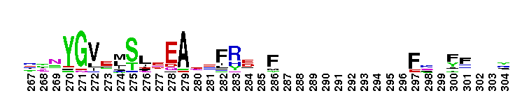
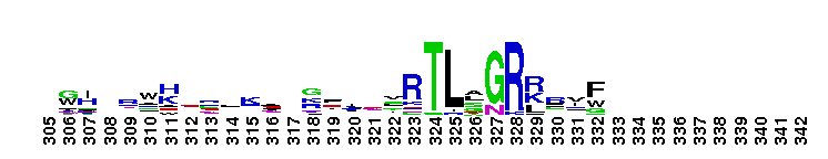
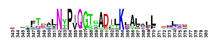

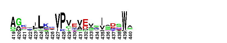
Weblogos are Copyright (c) 2002 Regents of the University of California
| DMDM_info@umbc.edu | 1000 Hilltop Circle, Baltimore, MD 21250 | Department of Biological Sciences | Phone: 410-455-2258 |