| ||||||||||||||||||||||


 Catalytic domain of metazoan phosphoinositide-specific phospholipase C-delta. This subfamily corresponds to the catalytic domain present in metazoan phosphoinositide-specific phospholipase C (PI-PLC, EC 3.1.4.11)-delta isozymes. PI-PLC is a signaling enzyme that hydrolyzes the membrane phospholipids phosphatidylinositol-4,5-bisphosphate (PIP2) to generate two important second messengers in eukaryotic signal transduction cascades, Inositol 1,4,5-trisphosphate (InsP3) and diacylglycerol (DAG). InsP3 triggers inflow of calcium from intracellular stores, while DAG, together with calcium, activates protein kinase C, which then phosphorylates other molecules, leading to altered cellular activity. Calcium is required for the catalysis. PLC-delta represents a class of mammalian PI-PLC that has an N-terminal pleckstrin homology (PH) domain, an array of EF hands, a PLC catalytic core domain, and a C-terminal C2 domain. This CD corresponds to the catalytic domain which is a TIM barrel with two highly conserved regions (X and Y) split by a highly degenerate linker sequence. There are three PI-PLC-delta isozymes (1,3 and 4). PI-PLC-delta1 is relatively well characterized. It is activated by high calcium levels generated by other PI-PLC family members, and therefore functions as a calcium amplifier within the cell. Different PI-PLC-delta isozymes have different tissue distribution and different subcellular locations. PI-PLC-delta1 is mostly a cytoplasmic protein, PI-PLC-delta3 is located in the membrane, and PI-PLC-delta4 is predominantly detected in the cell nucleus. Aside from three PI-PLC-delta isozymes identified in mammals, some eukaryotic PI-PLC-delta homologs have been classified to this CD.
 No pairwise interactions are available for this conserved domain.


Tips:  Range on the Protein: Protein ID Protein Position Domain Position: 

|
|---|


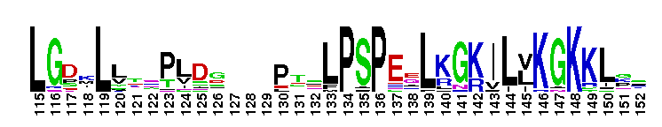


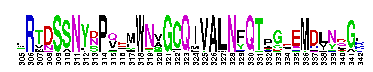

Weblogos are Copyright (c) 2002 Regents of the University of California
| DMDM_info@umbc.edu | 1000 Hilltop Circle, Baltimore, MD 21250 | Department of Biological Sciences | Phone: 410-455-2258 |