| ||||||||||||||||||||


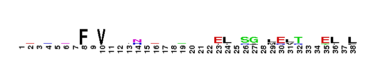
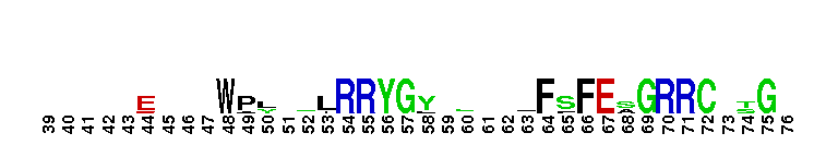
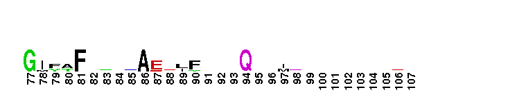
 Fibroblast growth factor receptor substrate 2 phosphotyrosine-binding domain. FRS2 (also called Suc1-associated neurotrophic factor (SNT)-induced tyrosine-phosphorylated target) proteins are membrane-anchored adaptor proteins. They are composed of an N-terminal myristoylation site followed by a phosphotyrosine binding (PTB) domain, which has a PH-like fold, and a C-terminal effector domain containing multiple tyrosine and serine/threonine phosphorylation site. The FRS2/SNT proteins show increased tyrosine phosphorylation by activated receptors, such as fibroblast growth factor receptor (FGFR) and TrkA, recruit SH2 domain containing proteins such as Grb2, and mediate signals from activated receptors to a variety of downstream pathways. The PTB domains of the SNT proteins directly interact with the canonical NPXpY motif of TrkA in a phosphorylationdependent manner, they directly bind to the juxtamembrane region of FGFR in a phosphorylation-independent manner. PTB domains have a common PH-like fold and are found in various eukaryotic signaling molecules. This domain was initially shown to binds peptides with a NPXY motif with differing requirements for phosphorylation of the tyrosine, although more recent studies have found that some types of PTB domains can bind to peptides lack tyrosine residues altogether. In contrast to SH2 domains, which recognize phosphotyrosine and adjacent carboxy-terminal residues, PTB-domain binding specificity is conferred by residues amino-terminal to the phosphotyrosine. PTB domains are classified into three groups: phosphotyrosine-dependent Shc-like, phosphotyrosine-dependent IRS-like, and phosphotyrosine-independent Dab-like PTB domains. This cd is part of the IRS-like subgroup.
 No pairwise interactions are available for this conserved domain.


Tips:  Range on the Protein: Protein ID Protein Position Domain Position: 

|
|---|


Weblogos are Copyright (c) 2002 Regents of the University of California
| DMDM_info@umbc.edu | 1000 Hilltop Circle, Baltimore, MD 21250 | Department of Biological Sciences | Phone: 410-455-2258 |