| |||||||||||||||||||


 Insulin receptor substrate phosphotyrosine-binding domain (PTBi). Insulin receptor substrate (IRS) molecules are mediators in insulin signaling and play a role in maintaining basic cellular functions such as growth and metabolism. They act as docking proteins between the insulin receptor and a complex network of intracellular signaling molecules containing Src homology 2 (SH2) domains. Four members (IRS-1, IRS-2, IRS-3, IRS-4) of this family have been identified that differ as to tissue distribution, subcellular localization, developmental expression, binding to the insulin receptor, and interaction with SH2 domain-containing proteins. IRS molecules have an N-terminal PH domain, followed by an IRS-like PTB domain which has a PH-like fold. PTB domains have a common PH-like fold and are found in various eukaryotic signaling molecules. This domain was initially shown to binds peptides with a NPXY motif with differing requirements for phosphorylation of the tyrosine, although more recent studies have found that some types of PTB domains can bind to peptides lack tyrosine residues altogether. In contrast to SH2 domains, which recognize phosphotyrosine and adjacent carboxy-terminal residues, PTB-domain binding specificity is conferred by residues amino-terminal to the phosphotyrosine. PTB domains are classified into three groups: phosphotyrosine-dependent Shc-like, phosphotyrosine-dependent IRS-like, and phosphotyrosine-independent Dab-like PTB domains. This cd is part of the IRS-like subgroup.
 No pairwise interactions are available for this conserved domain.


Tips:  Range on the Protein: Protein ID Protein Position Domain Position: 

|
|---|


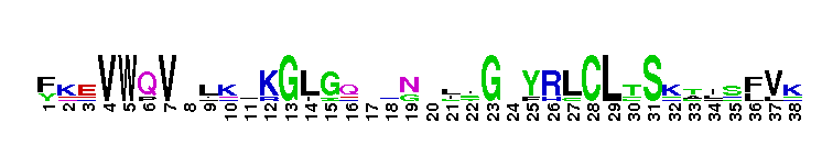
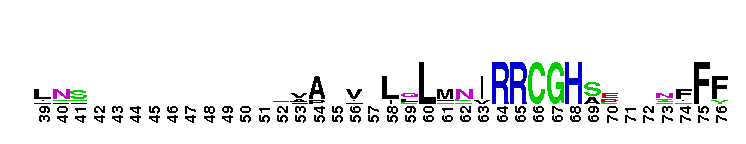
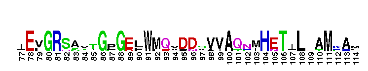

Weblogos are Copyright (c) 2002 Regents of the University of California
| DMDM_info@umbc.edu | 1000 Hilltop Circle, Baltimore, MD 21250 | Department of Biological Sciences | Phone: 410-455-2258 |