| ||||||||||||||||||||


 Adenylation domain of proteins similar to ATP-dependent polynucleotide ligases. ATP-dependent polynucleotide ligases catalyze the phosphodiester bond formation of nicked nucleic acid substrates using ATP as a cofactor in a three step reaction mechanism. This family includes ATP-dependent DNA and RNA ligases. DNA ligases play a vital role in the diverse processes of DNA replication, recombination and repair. ATP-dependent DNA ligases have a highly modular architecture, consisting of a unique arrangement of two or more discrete domains, including a DNA-binding domain, an adenylation or nucleotidyltransferase (NTase) domain, and an oligonucleotide/oligosaccharide binding (OB)-fold domain. The adenylation domain binds ATP and contains many active site residues. Together with the C-terminal OB-fold domain, it comprises a catalytic core unit that is common to most members of the ATP-dependent DNA ligase family. The catalytic core contains six conserved sequence motifs (I, III, IIIa, IV, V and VI) that define this family of related nucleotidyltransferases including eukaryotic GRP-dependent mRNA-capping enzymes. The catalytic core contains both the active site as well as many DNA-binding residues. The RNA circularization protein from archaea and bacteria contains the minimal catalytic unit, the adenylation domain, but does not contain an OB-fold domain. This family also includes the m3G-cap binding domain of snurportin, a nuclear import adaptor that binds m3G-capped spliceosomal U small nucleoproteins (snRNPs), but doesn't have enzymatic activity.
 No pairwise interactions are available for this conserved domain.

Tips:  Range on the Protein: Protein ID Protein Position Domain Position: 

|
|---|


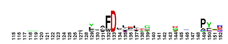

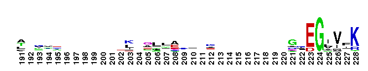

Weblogos are Copyright (c) 2002 Regents of the University of California
| DMDM_info@umbc.edu | 1000 Hilltop Circle, Baltimore, MD 21250 | Department of Biological Sciences | Phone: 410-455-2258 |