| |||||||||||||||||||


 Heme-copper oxidase subunit I. Heme-copper oxidases are transmembrane protein complexes in the respiratory chains of prokaryotes and mitochondria which catalyze the reduction of O2 and simultaneously pump protons across the membrane. The superfamily is diverse in terms of electron donors, subunit composition, and heme types. The number of subunits varies from three to five in bacteria and up to 13 in mammalian mitochondria. It has been proposed that Archaea acquired heme-copper oxidases through gene transfer from Gram-positive bacteria. Membership in the superfamily is defined by subunit I, which contains a heme-copper binuclear center (the active site where O2 is reduced to water) formed by a high-spin heme and a copper ion. It also contains a low-spin heme, believed to participate in the transfer of electrons to the binuclear center. Only subunit I is common to the entire superfamily. For every reduction of an O2 molecule, eight protons are taken from the inside aqueous compartment and four electrons are taken from the electron donor on the opposite side of the membrane. The four electrons and four of the protons are used in the reduction of O2; the four remaining protons are pumped across the membrane. This charge separation of four charges contributes to the electrochemical gradient used for ATP synthesis. Two proton channels, the D-pathway and K-pathway, leading to the binuclear center have been identified in subunit I of cytochrome c oxidase (CcO) and ubiquinol oxidase. A well-defined pathway for the transfer of pumped protons beyond the binuclear center has not been identified. Electron transfer occurs in two segments: from the electron donor to the low-spin heme, and from the low-spin heme to the binuclear center. The first segment can be a multi-step process and varies among the different families, while the second segment, a direct transfer, is consistent throughout the superfamily.
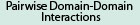 No pairwise interactions are available for this conserved domain.


Tips:  Range on the Protein: Protein ID Protein Position Domain Position: 

|
|---|


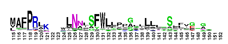


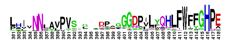


Weblogos are Copyright (c) 2002 Regents of the University of California
| DMDM_info@umbc.edu | 1000 Hilltop Circle, Baltimore, MD 21250 | Department of Biological Sciences | Phone: 410-455-2258 |