| |||||||||||||||||||||||||||||||||


 Third Ig domain of contactin. Ig3_Contactin_like: Third Ig domain of contactins. Contactins are neural cell adhesion molecules and are comprised of six Ig domains followed by four fibronectin type III(FnIII) domains anchored to the membrane by glycosylphosphatidylinositol. The first four Ig domains form the intermolecular binding fragment, which arranges as a compact U-shaped module via contacts between Ig domains 1 and 4, and between Ig domains 2 and 3. Contactin-2 (TAG-1, axonin-1) may play a part in the neuronal processes of neurite outgrowth, axon guidance and fasciculation, and neuronal migration. This group also includes contactin-1 and contactin-5. The different contactins show different expression patterns in the central nervous system. During development and in adulthood, contactin-2 is transiently expressed in subsets of central and peripheral neurons. Contactin-5 is expressed specifically in the rat postnatal nervous system, peaking at about 3 weeks postnatal, and a lack of contactin-5 (NB-2) results in an impairment of neuronal act ivity in the rat auditory system. Contactin-5 is highly expressed in the adult human brain in the occipital lobe and in the amygdala. Contactin-1 is differentially expressed in tumor tissues and may, through a RhoA mechanism, facilitate invasion and metastasis of human lung adenocarcinoma.
 No pairwise interactions are available for this conserved domain.


Tips:  Range on the Protein: Protein ID Protein Position Domain Position: 

|
|---|


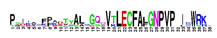
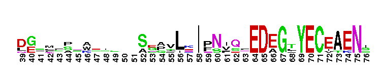
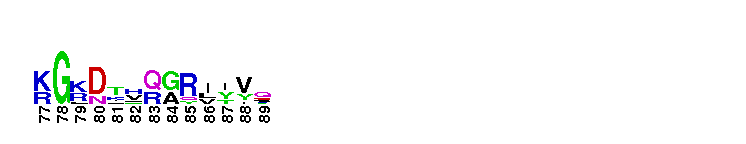
Weblogos are Copyright (c) 2002 Regents of the University of California
| DMDM_info@umbc.edu | 1000 Hilltop Circle, Baltimore, MD 21250 | Department of Biological Sciences | Phone: 410-455-2258 |