| |||||||||||||||||||||||||||


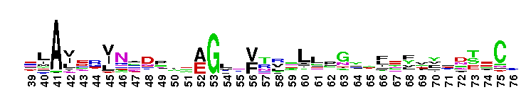
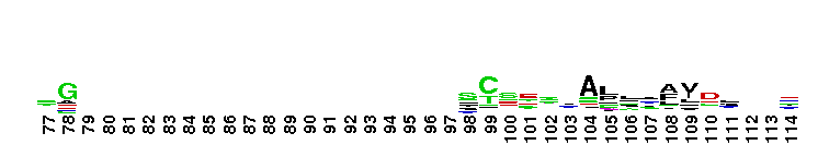
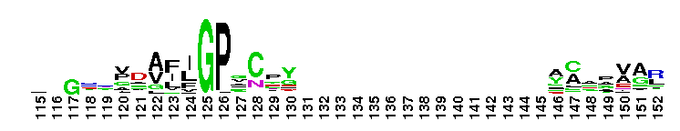
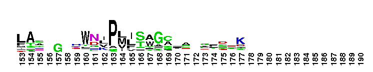
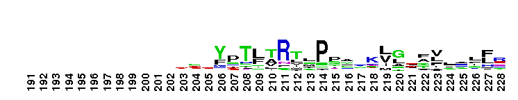
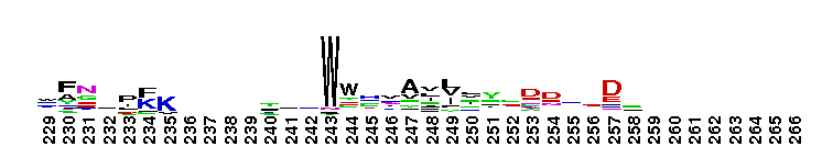
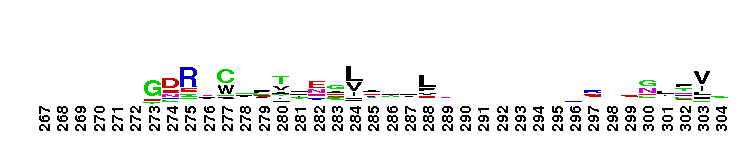
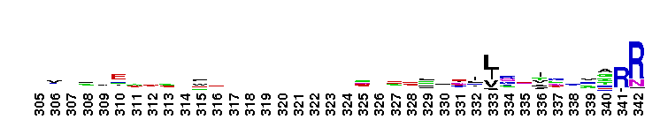
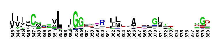
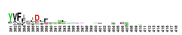
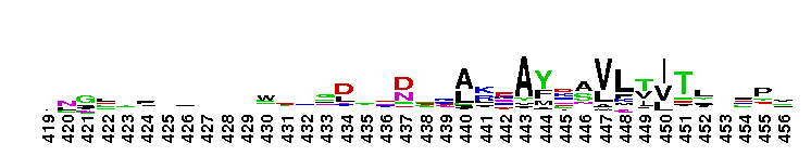
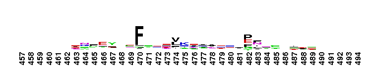
 Ligand-binding domain of membrane guanylyl-cyclase receptors. Ligand-binding domain of membrane guanylyl-cyclase receptors. Membrane guanylyl cyclases (GC) have a single membrane-spanning region and are activated by endogenous and exogenous peptides. This family can be divided into three major subfamilies: the natriuretic peptide receptors (NPRs), sensory organ-specific membrane GCs, and the enterotoxin/guanylin receptors. The binding of peptide ligands to the receptor results in the activation of the cytosolic catalytic domain. Three types of NPRs have been cloned from mammalian tissues: NPR-A/GC-A, NPR-B/ GC-B, and NPR-C. In addition, two of the GCs, GC-D and GC-G, appear to be pseudogenes in humans. Atrial natriuretic peptide (ANP) and brain natriuretic peptide (BNP) are produced in the heart, and both bind to the NPR-A. NPR-C, also termed the clearance receptor, binds each of the natriuretic peptides and can alter circulating levels of these peptides. The ligand binding domain of the NPRs exhibits strong structural similarity to the type I periplasmic binding fold protein family.
 No pairwise interactions are available for this conserved domain.


Tips:  Range on the Protein: Protein ID Protein Position Domain Position: 

|
|---|


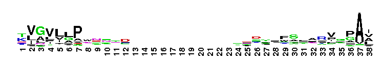
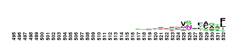
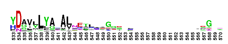
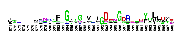
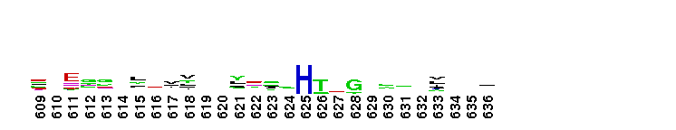
Weblogos are Copyright (c) 2002 Regents of the University of California
| DMDM_info@umbc.edu | 1000 Hilltop Circle, Baltimore, MD 21250 | Department of Biological Sciences | Phone: 410-455-2258 |