| |||||||||||||||||||


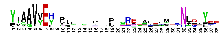
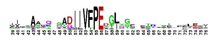
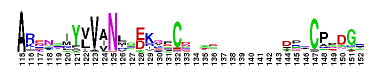
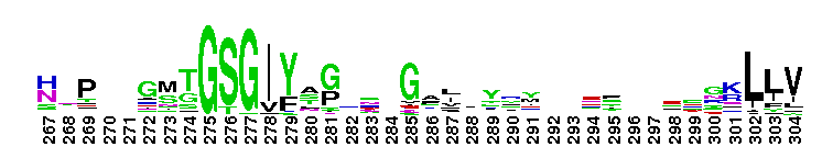
 biotinidase and vanins (class 4 nitrilases). These secondary amidases participate in vitamin recycling. Biotinidase (EC 3.5.1.12) has both a hydrolase and a transferase activity. It hydrolyzes free biocytin or small biotinyl-peptides produced during the proteolytic degradation of biotin-dependent carboxylases, to release free biotin (vitamin H), and it can transfer biotin to acceptor molecules such as histones. Biotinidase deficiency in humans is an autosomal recessive disorder characterized by neurological and cutaneous symptoms. This subgroup includes the three human vanins, vanin1-3. Vanins are ectoenzymes, Vanin-1, and -2 are membrane associated, vanin-3 is secreted. They are pantotheinases (EC 3.5.1.92, pantetheine hydrolase), which convert pantetheine, to pantothenic acid (vitamin B5) and cysteamine (2-aminoethanethiol, a potent anti-oxidant). They are potential targets for therapeutic intervention in inflammatory disorders. Vanin-1 deficient mice lacking free cysteamine are less susceptible to intestinal inflammation, and expression of vanin-1 and -3 is induced as part of the inflammatory-regenerative differentiation program of human epidermis. This subgroup belongs to a larger nitrilase superfamily comprised of nitrile- or amide-hydrolyzing enzymes and amide-condensing enzymes, which depend on a Glu-Lys-Cys catalytic triad. This superfamily has been classified in the literature based on global and structure based sequence analysis into thirteen different enzyme classes (referred to as 1-13), this subgroup corresponds to class 4. Members of this superfamily generally form homomeric complexes, the basic building block of which is a homodimer.
 No pairwise interactions are available for this conserved domain.


Tips:  Range on the Protein: Protein ID Protein Position Domain Position: 

|
|---|


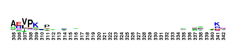
Weblogos are Copyright (c) 2002 Regents of the University of California
| DMDM_info@umbc.edu | 1000 Hilltop Circle, Baltimore, MD 21250 | Department of Biological Sciences | Phone: 410-455-2258 |