| |||||||||||||||||||


 Glyoxalase I catalyzes the isomerization of the hemithioacetal, formed by a 2-oxoaldehyde and glutathione, to S-D-lactoylglutathione. Glyoxalase I (also known as lactoylglutathione lyase; EC 4.4.1.5) is part of the glyoxalase system, a two-step system for detoxifying methylglyoxal, a side product of glycolysis. This system is responsible for the conversion of reactive, acyclic alpha-oxoaldehydes into the corresponding alpha-hydroxyacids and involves 2 enzymes, glyoxalase I and II. Glyoxalase I catalyses an intramolecular redox reaction of the hemithioacetal (formed from methylglyoxal and glutathione) to form the thioester, S-D-lactoylglutathione. This reaction involves the transfer of two hydrogen atoms from C1 to C2 of the methylglyoxal, and proceeds via an ene-diol intermediate. Glyoxalase I has a requirement for bound metal ions for catalysis. Eukaryotic glyoxalase I prefers the divalent cation zinc as cofactor, whereas Escherichia coil and other prokaryotic glyoxalase I uses nickel. However, eukaryotic Trypanosomatid parasites also use nickel as a cofactor, which could possibly be explained by acquiring their GLOI gene by horizontal gene transfer. Human glyoxalase I is a two-domain enzyme and it has the structure of a domain-swapped dimer with two active sites located at the dimer interface. In yeast, in various plants, insects and Plasmodia, glyoxalase I is four-domain, possibly the result of a further gene duplication and an additional gene fusing event.
 No pairwise interactions are available for this conserved domain.


Tips:  Range on the Protein: Protein ID Protein Position Domain Position: 

|
|---|


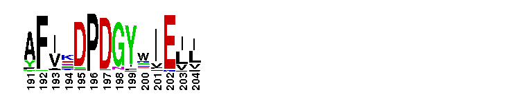
Weblogos are Copyright (c) 2002 Regents of the University of California
| DMDM_info@umbc.edu | 1000 Hilltop Circle, Baltimore, MD 21250 | Department of Biological Sciences | Phone: 410-455-2258 |