| |||||||||||||||||||||


 First immunoglobulin (Ig)-like domain found in Leukocyte Ig-like receptors (LILR)B1 (also known as LIR-1) and similar proteins. Ig1_LILRB1_like: domain similar to the first immunoglobulin (Ig)-like domain found in Leukocyte Ig-like receptors (LILR)B1 (also known as LIR-1). This group includes, LILRA5 (LIR9), an activating natural cytotoxicity receptor NKp46, and the immune-type receptor glycoprotein VI (GPVI). LILRs are a family of immunoreceptors expressed on expressed on T and B cells, on monocytes, dendritic cells, and subgroups of natural killer (NK) cells. The human LILR family contains nine proteins (LILRA1-3,and 5, and LILRB1-5). From functional assays, and as the cytoplasmic domains of various LILRs, for example LILRB1 (LIR-1), LILRB2 (LIR-2), and LILRB3 (LIR-3) contain immunoreceptor tyrosine-based inhibitory motifs (ITIMs) it is thought that LIR proteins are inhibitory receptors. Of the eight LIR family proteins, only LIR-1(LILRB1), and LIR-2 (LILRB2), show detectable binding to class I MHC molecules; ligands for the other members have yet to be determined. The extracellular portions of the different LIR proteins contain different numbers of Ig-like domains for example, four in the case of LILRB1 (LIR-1), and LILRB2 (LIR-2), and two in the case of LILRB4 (LIR-5). The activating natural cytotoxicity receptor NKp46. is expressed in natural killer cells, and is organized as an extracellular portion having two Ig-like extracellular domains, a transmembrane domain, and a small cytoplasmic portion. GPVI, which also contains two Ig-like domains, participates in the processes of collagen-mediated platelet activation and arterial thrombus formation.
 No pairwise interactions are available for this conserved domain.


Tips:  Range on the Protein: Protein ID Protein Position Domain Position: 

|
|---|


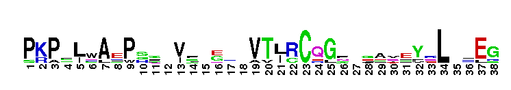
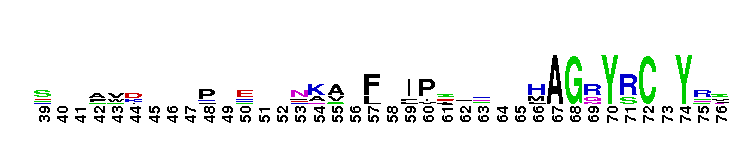

Weblogos are Copyright (c) 2002 Regents of the University of California
| DMDM_info@umbc.edu | 1000 Hilltop Circle, Baltimore, MD 21250 | Department of Biological Sciences | Phone: 410-455-2258 |