| ||||||||||||||||||||||||||||


 The ligand binding domain of REV-ERB receptors, members of the nuclear receptor superfamily. The ligand binding domain (LBD) of REV-ERB receptors: REV-ERBs are transcriptional regulators belonging to the nuclear receptor superfamily. They regulate a number of physiological functions including the circadian rhythm, lipid metabolism, and cellular differentiation. The LBD domain of REV-ERB is unusual in the nuclear receptor family by lacking the AF-2 region that is responsible for coactivator interaction. REV-ERBs act as constitutive repressors because of their inability to bind coactivators. REV-ERB receptors can bind to two classes of DNA response elements as either a monomer or heterodimer, indicating functional diversity. When bound to the DNA, they recruit corepressors (NcoR/histone deacetylase 3) to the promoter, resulting in repression of the target gene. The porphyrin heme has been demonstrated to function as a ligand for REV-ERB. Like other members of the nuclear receptor (NR) superfamily of ligand-activated transcription factors, REV-ERB receptors have a central well conserved DNA binding domain (DBD), a variable N-terminal domain, a non-conserved hinge and a C-terminal ligand binding domain (LBD).
 No pairwise interactions are available for this conserved domain.


Tips:  Range on the Protein: Protein ID Protein Position Domain Position: 

|
|---|


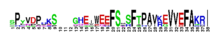
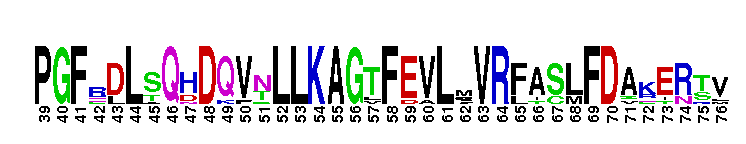
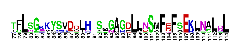
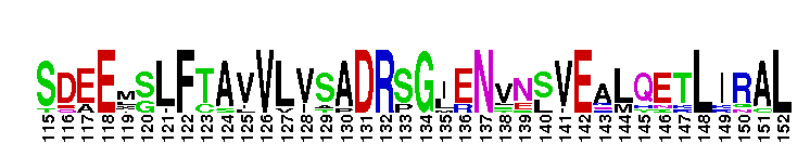

Weblogos are Copyright (c) 2002 Regents of the University of California
| DMDM_info@umbc.edu | 1000 Hilltop Circle, Baltimore, MD 21250 | Department of Biological Sciences | Phone: 410-455-2258 |